
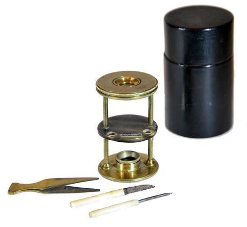
Withering-type botanical microscope, 1780
The “Withering-type Microscope” is named for its inventor, Dr. William Withering (1741-1799), an English physician and botanist who graduated with a degree in medicine 1766 in Edinburgh. Inspired by the taxonomical work and systematic classification of Carl Linnæus (1707-1778), Withering (1776) applied the Linnaean taxonomical system of classification to British plants in a seminal, two volume work, A Botanical arrangement of all the vegetables naturally growing in the British Isles. The earliest reference to a small botanical microscope of Withering’s design appeared in the first edition of this book. There, Withering indicated this microscope was developed for field dissections of flowers and other plant parts. While there is no surviving example of this exact design, close relatives of this type do exist, made either completely of brass or of ivory with brass pillars. Ivory models can be tentatively dated to 1776-1785, as by 1787 a newer model with a hollowed stage in an all-brass configuration already predominated. In turn, it was preceded by the brief appearance of a transitional brass model but with solid stage of ivory or horn (seen here). This version is extremely rare and must have been produced in very small numbers. By 1787 all these varieties were not recorded anymore in the literature.
References: SML: A242712; Goren 2014.
References: SML: A242712; Goren 2014.
Prof. Yuval Goren's Collection of the History of the Microscope
Chapter 12: The Aquatic Microscope

© Microscope History all rights reserved
Early Aquatic Microscope, ~1750
The 'Aquatic microscope' is a design originally suggested by the Swiss naturalist Abraham Trembley (1710-1784) for his groundbreaking study of the hydra. This concept was later brought to life by microscope maker John Cuff (circa 1708-1772) for the Irish naturalist John Ellis (1707-1776). Ellis was highly esteemed by Carl Linnaeus (1707-1778), who regarded him as one of the brightest stars in natural history. It is interesting to note that Linnaeus took an identical instrument with him on his travels. This instrument was made in London by John Cuff around 1750. Ellis was elected a Fellow of the Royal Society (FRS) in 1754. In 1755, Ellis published his influential work on corals, which was translated into French the following year, earning him international acclaim.
Ellis sought a highly portable microscope that would allow him to easily observe the activities of the hydra and other small water organisms placed on a watch glass on the microscope's stage. The first model was constructed by Cuff for Ellis in 1752. The design gained popularity, leading to the production of various forms of this microscope across England and Europe. Over time, improvements to the design included rack-and-pinion focusing, replacing the simpler push-fit focusing mechanism found in the original models. Additionally, Raspail modified the design by converting the arm's forward and backward motion carrying the eyepiece into a fine motion controlled by a screw mechanism.
In 1832, Charles Darwin took an advanced version of the Aquatic microscope, made by Bancks of London (which is also represented in this collection), on his famous voyage aboard HMS Beagle.
This early model of the aquatic microscope is mounted in a wooden case covered with ray skin. It features a central mount for the pillar, Lieberkuhn lenses of various magnifications that can be attached to a bar above the circular stage, and a convex mirror located below.


This early version of an aquatic microscope is housed in a wooden case coated with ray skin. It features a central mount for the pillar and interchangeable Lieberkuhn lenses of varying magnifications, which can be attached to a bar above the circular stage. Below the stage is a specimen holder and a convex mirror. The inner red lining of the case is characteristic of the microscope makers Benjamin Martin or John Cuff.

Early Aquatic Microscope by Barthelon a Marseille, France, ~1750
During the 18th century, several scientific instruments were invented and became quite successful. One notable example is the 'Aquatic Microscope,' developed by John Cuff and other manufacturers in London. This design was soon replicated by prominent microscope producers across continental Europe. These replicas often featured modifications and improvements, as the copying manufacturers were not held to the same standards as the English optical and instrument makers.
The microscope in question is an early replica of John Cuff's original design for John Ellis in London during the mid-18th century. This improved model includes a thinner box, a more comfortable arrangement of internal components, enhanced connectivity of the stage tweezers, and decorative embellishments on the stage. However, it generally remains true to Cuff's original design. In this design, the rod that supports the lens carrier enters a tube attached to the pole, which is connected to the mirror, stage, and lens system, rather than fitting into a hole in the center.

The inscription on the central stand of the microscope reads "BARTHELON · A · MARSEILLE." However, this name is unfamiliar to us, as there were no known manufacturers or retailers associated with it in France during that time period. It is possible that this name belongs to the microscope's owner; however, it was uncommon for owners to sign their devices in the 18th century. However, in this collection this practice is represented also by an earlier compound microscope, clearly of Italian make but signed by a French owner from Rouen.
References: MHS: 42845; Turner 1989: 270-1; Billings: P. 158, Fig. 297, AFIP 49163-60-4713-37; Whipple: 1824; Harvard Univ. 1007; Mus. Galileo 3212;