
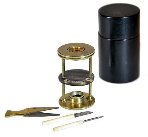
Withering-type botanical microscope, 1780
The “Withering-type Microscope” is named for its inventor, Dr. William Withering (1741-1799), an English physician and botanist who graduated with a degree in medicine 1766 in Edinburgh. Inspired by the taxonomical work and systematic classification of Carl Linnæus (1707-1778), Withering (1776) applied the Linnaean taxonomical system of classification to British plants in a seminal, two volume work, A Botanical arrangement of all the vegetables naturally growing in the British Isles. The earliest reference to a small botanical microscope of Withering’s design appeared in the first edition of this book. There, Withering indicated this microscope was developed for field dissections of flowers and other plant parts. While there is no surviving example of this exact design, close relatives of this type do exist, made either completely of brass or of ivory with brass pillars. Ivory models can be tentatively dated to 1776-1785, as by 1787 a newer model with a hollowed stage in an all-brass configuration already predominated. In turn, it was preceded by the brief appearance of a transitional brass model but with solid stage of ivory or horn (seen here). This version is extremely rare and must have been produced in very small numbers. By 1787 all these varieties were not recorded anymore in the literature.
References: SML: A242712; Goren 2014.
References: SML: A242712; Goren 2014.
Prof. Yuval Goren's Collection of the History of the Microscope

Carlo Antonio Tortoni, screw-barrel compound microscope, 1683
Carlo Antonio Tortoni (1640-1700) was a priest, physicist, and mathematician born in Recanati, near Ancona, in the Papal Marches. After being appointed chamberlain at the court of Pope Alexander VII, he moved to Rome, where he lived during the last decades of the seventeenth century. Aside from his interest in the sciences and the development of scientific instruments of his time, little else is known about him. In Rome, Tortoni joined the Physico-Mathematical Academy (Accademia Fisico-Matematica), which was established in 1677 under the patronage of Queen Christina of Sweden (1626-1689).
Tortoni was an active member of the Accademia. Among the various publications sponsored by it, two notable pamphlets on microscopes were published in 1687 in Rome. One pamphlet, titled "Instructions for the Two Types of Tortoni Microscopes Newly Invented and Brought to Light," described two screwbarrel microscopes a design now commonly accepted to be invented by Tortoni. The second publication was a public letter from Tortoni, which included new insights presented by Giovanni Battista Vacondio, entitled: "Letter in which the Prerogative of the Said Microscope is Mentioned, Along with Its Composition, Numerous Experiments Conducted, the Demonstration of the Parabolic Figure, and Various Designs of Enlargement that the Instrument Provides." These instructional pamphlets accompanied one of Tortoni’s two models of his screwbarrel microscope.
Tortoni's first model, which is shown here, is this portable handheld screwbarrel microscope of 1683, which he demonstrated in the palace of Giovanni Ciampini (1633-1698), pioneer in archaeology, on August 5, 1685. The screw served three key purposes: it facilitated focus adjustment for observed objects, allowed for a range of magnifications by changing the focal length, and enabled the instrument to function as a telescope. Tortoni provided a partial description of his microscope in a publication but omitted the technical details due to concerns about plagiarism. Consequently, he shared only certain aspects of the instrument rather than the complete design, which can be observed by comparing the figures with the entire apparatus as shown below. Due to poor health, he was unable to document his microscopes as he wished and was reluctant to reveal his studies until they were fully developed, first on paper and then in practice.
© Microscope History all rights reserved


© Microscope History all rights reserved
Quickly, his fellow countrymen began to replicate the "screw-barrel" principle in their microscope designs, adding innovative features. Notably, Filippo Bonanni (or Buonanni, 1638-1725) introduced a spring mechanism that pushed the slider toward the frame holding the object to be observed, ensuring it aligned with the fixed ocular lens. It wasn't until the early twentieth century that Tortoni was officially recognized as the true inventor of this principle.


The value attributed to Tortoni for his innovative techniques is evident in his expertise and meticulous approach to describing his observations through microscopy. He remarked on the shapes and movements of the micro-organisms he studied, stating: "...in cases of gangrene, the majority of those observed could be a mine of worms, as well as in sores that contained varying quantities of animals in different colors, some with black heads. In human blood, one can see continuous changes resembling brilliant little diamonds, some in conical shapes, while others look like small seeds of changing garnets. In febrile blood, pieces of vermilion watermelon can still be recognized, along with areas of ash color that contain small, worm-like creatures of citron color. In cases of malignant fevers, the blood is filled with black points, which knowledgeable doctors say nature transmits to the surface of the skin, commonly referred to as petechiae."
In addition to inventing the screw-barrel microscope, Tortoni should also be remembered for his pioneering use of microscopy in medical experimentation. His versatility is evident in another of his publications, which addresses a topic seemingly far removed from his microscope designes: the creation of a therapeutic balm. Tortoni ventured into this new field, captivated by the miniscule world he observed through his instruments. In this world, minute creatures and other unidentified entities exhibit various behaviors depending on their environments.
Impressed by his microscopical observations, Tortoni recognized the need for new remedies to address what he believed were the causes of many diseases. As a result, he developed a balm he published as Balsamo Tortoriano, which he claimed could miraculously cure numerous ailments. He detailed his findings in a publication dated 1689. However, the exact composition of the balm remains unknown, as Tortoni closely guarded it out of mistrust and fear of plagiarism.
Portrait of Carlo Antonio Tortoni, from Istruzione delle due sorti di microscopii tortoniani nuovamente inventati (Rome: 1687) Houghton Library, Harvard University (public domain).

© Microscope History all rights reserved
To the best of our knowledge, only two examples of this microscope have survived. One, designated No. 238, is presented in the Kern Collection of historical scientific instruments. It is made of boxwood but does not have a case. The second example, which is the one displayed here, is made from ivory and comes in a fitted wooden case. This case features inner chamois lining and compartments for two ivory sliders, along with special brass tweezers for handling them. Additionally, the outer cover is coated with hide and is hot-stamped with motifs of the Florentine (Medici) fleur-de-lys. It also has an iron-hinged top equipped with a lock.
This suggests that this microscope was part of the collection of scientific instruments held by Cosimo III de' Medici (1642 – 1723), Grand Duke of Tuscany from 1670 until his death in 1723.

© Microscope History all rights reserved
Medici Grand Dukes as Patrons of Science
The extensive correspondence found in the Medici Grand Ducal archives, dating from 1537 to 1743, highlight the Medici family's significant interest in various fields of natural and applied sciences. These fields included hydraulics, engineering, botany, human and veterinary anatomy, pharmacology, metallurgy, mineralogy, cartography, meteorology, astrology, astronomy, mathematics, optics, and alchemy.
The Medici's patronage of scientific studies, scientists, and technological advancements was rooted in a fundamental belief: that knowledge of science and mastery of technology would enhance their political power. They were interested in how scientific discoveries and applications could improve everyday life, commerce, and communication.
The Medici supported significant figures such as Luca Ghini in botany, Andreas Vesalius in anatomy, Galileo Galilei in astronomy and mathematics, Nicolas Steno in anatomy and geology, and Francesco Redi in medicine. They contributed to various educational institutions, teaching hospitals like Santa Maria Nuova in Florence, academies such as the Accademia del Cimento in Florence, and botanical gardens like the Orto Botanico in Pisa and the Giardino dei Semplici in Florence. They also played a role in the publication of both ancient and modern scientific works.
Cosimo I had a particular interest in acquiring the secrets of glassmaking, sugar refining, and porcelain production. Ferdinand began assembling a collection of scientific artifacts and instruments, while Cosimo II showed an affinity for nautical engineering.
Over the years, the Medici Family, renowned patrons of art and science, assembled an impressive collection of scientific instruments. It began with Cosimo I, the founder of the Grand Duchy of Tuscany. His sons and successors further enriched it: Francesco I, who focused mainly on natural history and alchemical collections; and Ferdinando I, who acquired numerous mathematical, nautical, and cosmographical instruments. Cosimo II had the distinction of adding Galileo's groundbreaking instruments to the collection. Later, beautifully crafted glass thermometers were produced in the glassworks of Palazzo Pitti for the Accademia del Cimento, which was established by Grand Duke Ferdinando II and Prince Leopoldo de' Medici. A notable figure among the later Medici rulers is Cosimo III, who was a patron of the mathematician Vincenzo Viviani, Galileo's last disciple.
Balms and the Medici court
The Medici family of Florence, first rose to power in the fourteenth century, primarily through commerce and banking. Their influence allowed them to maintain power for nearly three centuries. The family supported the rise of four popes, which granted them significant influence throughout Christendom and expanded their reach beyond Florence. The remains of family members are interred in the San Lorenzo Basilica, where the Medici Chapels are located. These remains have been studied as part of a research project. Since the project's inception, several studies have investigated the health of the family over time, while additional research has explored the embalming and autopsy techniques used in Italy during that period. Entomological and arachnological examinations of the contents of the embalming jars have also been previously published.
One study focuses on recovering pollen from an embalming jar found within the San Lorenzo architectural complex. The pollen grains are some of the most intriguing discoveries from the jar. Various samples of human viscera fragments, sponges, and cloth were collected from embalming jars belonging to members of the Medici family. One jar was labeled with the name of Vittoria della Rovere, who died in March 1694. This jar contained viscera fragments identified as a section of a collapsed intestine.
Analysis revealed that the intestine sample from Vittoria della Rovere contained a large concentration of pollen from the Myrtaceae family. The Myrtaceae pollen was sometimes observed in clusters during analysis, suggesting purposeful ingestion of flowers, buds, or a substance derived from floral structures. Therefore, the high concentrations and clustering of Myrtaceae pollen grains recovered from this sample likely reflect dietary or medicinal practices. Scanning electron microscopy indicated that the pollen came from cloves (Syzygium aromaticum). It is most likely that Vittoria della Rovere consumed cloves for medicinal or culinary purposes shortly before her death.
Was this the Balsamo Tortoriano? We may never know.
In 1670, at the age of twenty-eight, Cosimo de' Medici inherited the Grand Dukedom of Tuscany from his father, Ferdinando II de' Medici. He ruled for over fifty years as Cosimo III. Although his personal and political life could hardly be considered successful, his support for the arts and sciences somewhat compensated for his inability to reverse the decline of the state that had begun during his father's reign. During this period, the arts, sciences, and even religion were deeply interconnected, with every educated individual often acting as a sort of pantologist, exploring a wide range of subjects. This integration was partly due to the study of the seven liberal arts, a curriculum established in classical Rome that became the foundation for university education from the 12th century onward. Students would spend six years studying these subjects—grammar, logic, rhetoric, geometry, arithmetic, astronomy, and music—to earn a master’s degree.

© Microscope History all rights reserved
.jpg)
Cosimo III de' Medici, Grand Duke of Tuscany (public domain)

17th-century medicine in Italy and the microscope
During the conservation of the microscope case by Mr. Michael Maggen, he observed that it originally had a carrying strap, which made the small instrument highly portable. This portability has historical significance in the context of 17th-century Italy and Tortoni's experiments with infectious diseases. These experiments likely related to "animalcules" (i.e., microorganisms) found in the air and the effects of remedies developed to combat them.
Makers of scientific instruments in Rome often served the four Medici princes in Florence: Grand Duke Ferdinand II and his three brothers. In 1630, when the plague ravaged Tuscany, most upper-class members fled the city. In contrast, the Grand Duke and his brothers chose to stay in Florence, working tirelessly to care for the sick and provide as much assistance as possible. As a disciple of Galileo, Ferdinand dedicated much of his time to experimenting with scientific instruments.
During the 17th-century plague outbreak in Italy, the microscope emerged as a promising tool for scientific discovery. In Rome, the German Jesuit polymath Athanasius Kircher (1602–1680) was among the first to use it to study the plague, examining blood and publishing his findings. In 1653, French physician Pierre Borel described blood under a microscope, noting tiny creatures he likened to dolphins swimming in a "red ocean." He provided numerous observations, demonstrating how the microscope could help diagnose illnesses by revealing previously invisible symptoms. By early 1658, Kircher completed the "Examination of Plague", the first treatise to base conclusions on microscopical observations, dedicating it to Pope Alexander VII amid the devastation caused by the epidemic. Kircher described using a simple microscope gifted by Cardinal Giancarlo de' Medici to magnify various specimens. His earlier work foreshadowed the medical applications of the microscope, which Borel acknowledged in his observations. In 1657, Gaspar Schott, Kircher's disciple, published accounts of their microscopical experiments, which he hailed as groundbreaking. Schott mentioned Eustachio Divini, a prominent microscope maker in Rome, as a potential source for Kircher's instruments, emphasizing the community's efforts to advance microscopic research in response to the ongoing plague.
Other early microscope makers in the Italian sphere took this general idea. Giuseppe Campani (1635-1715) from Rome was one of Italy's most prominent optics manufacturers in the late 17th century. Over fifty years of activity, Campani produced many optical instruments with lenses of excellent workmanship. He included Bonanni's design while adding a second threading system of the draw-tube into the objective tube that provides the microscope with various magnifications. He even published it to perform microscopical examinations during medical and surgical activities. The idea was also copied by other manufacturers in the Italian countries, such as Pietro Patroni (1676/7-1744), François De Baillou (ca. 1700-1774), and others. Microscopes of a similar design (below) are also known with the signatures of some French makers or users.

Giuseppe Campani’s (1635-1715) compound microscopes designed for medical diagnosis. Special Collections, Stanford University Library
References
-
Bendini, S. 2021. "Chapter 20: Fleas Large as a Man’s Fist” (1686–1700). In: Giuseppe Campani, “Inventor Romae,” an Uncommon Genius (Nuncius Series, Vol. 8), Pp. 644–690. The Hauge.
-
Borel, P. Historiarum et observationum medico-physicarum Centuria IV (Paris, 1657 ed.; 1653), p. 198.
-
Buonanni, F. 1691. Observationes circa Viventia, quae in rebus non Viventibus reperiuntur. Cum Micrographia curiosa. Roma.
-
Clay, R. & Court, T. 1932. The History of the Microscope. London.
-
Di Napoli, C. Nuove inventioni di tubi ottici: dimostrate nell’Accademia fisicomatematica romana l’anno 1686 (In Roma: Nella Stamperia di Gio: Giacomo Komarek Boemo, 1686), 5; Novum microscopium authore Carolo Antonio Tortono, sacerdote piceno, Roma degente, Acta Eruditorum Octobris, no. 10 (1685): 478–80.
-
Didier F. 1999. Pierre Borel médecin et savant castrais du XVIIe siècle: textes choisis, trans. Jean Golfin and Foucault, Cahiers d’histoire du Centre d’étude d’histoire de la medicine de Toulouse, 7 (Toulouse: Pierre-C. Lile, 1999).
-
Findlen, P. 2022. Microscopic musings: Athanasius Kircher and the Roman plague of 1656–57. Harvard Library Bulletin, https://harvardlibrarybulletin.org/microscopic-musings-athanasius-kircher-and-roman-plague
-
Kircher, A. 1659. Athanasii Kircheri e Soc. Iesu scrutinium physico-medicum contagiosae luis, quae pestis dicitur ; quo origo, causae, signa, prognostica pestis, nec non insolentes malignantis naturae effectus, qui statis temporibus, caelestium influxuum virtute & efficacia, tum in elementis : tum in epidemijs hominum animantiumque morbis elucescunt, unà cum appropriatis remediorum antidotis nouâ doctrinâ in lucem eruuntur. Vorwort von Chr. Lang. Leipzig. https://curiosity.lib.harvard.edu/contagion/catalog/36-990058668780203941
-
Lippi, D. 2017. Early use of the microscope. The Lancet 389: 1793-1794. https://www.thelancet.com/journals/lancet/article/PIIS0140-6736%2817%2930049-1/fulltext
-
Reinhard, K. et al. 2018. Pollen evidence of medicine from an embalming jar associated with Vittoria della Rovere, Florence, Italy. Journal of Archaeological Science: Reports 21: 238-242. https://doi.org/10.1016/j.jasrep.2018.06.039
-
Riva, M. A., Borghi, L., Pagni, F. 2016. The first recorded use of microscopy in medicine: Pope Innocent XII's autopsy report. The Lancet 388: 559. DOI: 10.1016/S0140-6736(16)31210-7
-
Riva, M.A., Buontempo, V. 2025. Carlo Tortoni (1640–1700) and the early use of microscope in medical experimentation. Internal and Emergency Medicine 20: 323–325. https://doi.org/10.1007/s11739-024-03642-3
-
Tortoni, C. A. 1687. Lettera scritta da D. Carlo Antonio Tortoni Sac. All’Illustriss. Reverendiss. E Dottiss. Sig. Il Sig. D. Girolamo Ambrogio de Langenmantel Dottore dell’uno, e l’altra Legge, e Canonico Meritissimo nella Collegiata di S. Nauritzio in Augusta. Di nuove data in luce Da Gio. Battista Vacondio, Dottore dell’una, e l’altra Legge. Nella quale si accennano le prerogative del detto Microscopio, e sua composizione, con molte sperienze fatte, con la dimostrazione della figure Parabolica, e con alcuni disegni de gli ingrandimenti, che porta il medesimo. Roma: Gio. Giacomo Komarek.

Other early microscope makers in the Italian sphere took this general idea. Giuseppe Campani (1635-1715) from Rome was one of Italy's most prominent optics manufacturers in the late 17th century. Over fifty years of activity, Campani produced many optical instruments with lenses of excellent workmanship. He included Bonanni's design while adding a second threading system of the draw-tube into the objective tube that provides the microscope with various magnifications. He even published it to perform microscopical examinations during medical and surgical activities. The idea was also copied by other manufacturers in the Italian countries, such as Pietro Patroni (1676/7-1744), François De Baillou (ca. 1700-1774), and others. Microscopes of a similar design (below) are also known with the signatures of some French makers or users.
© Microscope History all rights reserved
Campani microscope signed Nicolas Dufur ARouen, ca. 1680-1700
Filippo Bonanni (or Buonanni, 1638-1725), a Jesuit scholar from Rome who worked in several scientific and technical fields, described in his book Micrographia Curiosa in 1691 a new type of compound microscope. The two types of compound microscopes he designed were significant innovations in the optics and mechanics of the late 17th-century microscope. One of them was intended for observation in transparent samples on slides or opaque surfaces in reflected light, based on a focusing mechanism in which the lens tube was screwed up and down inside the cylindrical body of the microscope in a configuration later called a screw-barrel, a configuration now commonly attributed to Carlo Antonio Tortoni. This configuration soon became the accepted method in one of the most popular single microscope models from the late 17th century and most of the 18th century.
Bonani entered the Jesuit Society in 1654 at the age of 17. In 1656 he was sent to study at the Society's prestigious Roman College as a student of Athanasius Kircher, working on producing microscope lenses. He used his lenses to build his microscope and develop scientific studies on multiple specimens. He also became a skilled engraver.
After Kircher's resignation from the position of professor of mathematics at the Roman College, Bonani was elected to succeed him. After Kircher's death in 1698, Bonani was appointed treasurer of the well-known Wunderkammer collected by Kircher and set up in the Roman College. He published a catalog of the collection entitled Musæum Kicherianum in 1709.
As a believer in the theories of spontaneous generation, he became bitter a critique of Francesco Redi's experimental work, defending the Aristotelian view. Although he raised important questions - such as whether observers through a microscope saw what they expected rather than what was there, later writers tended to underestimate him.


© Microscope History all rights reserved
In 1653, a French physician and chemist named Pierre Borel made the first recorded observation of blood under a microscope. He described seeing "animals in the shape of dolphins or whales" swimming in human blood, "as if in a red ocean." Over the next few years, he made other observations of bodily fluids under the microscope, such as the white corpuscles in serum and chyle that are not found in urine, as well as the valves of pores, vessels, and veins. Borel's work in anatomy paved the way for future scientific discoveries using microscopes.

© Microscope History all rights reserved
The microscope seen here has an engraved signature on the brass collar:
>>>>>>>>NICOLAS DUFUR AROUEN<<<<<<<<
The family name Dufur (or Dufour) is a French nickname for a baker, derived from "du four", which literally translates to "of the oven". It can also be a habitational name for someone from Le Four, which is the name of several places in various parts of France. However, there is no evidence of a manufacturer of scientific instruments with this name in Rouen or anywhere in France during the 17th or 18th centuries. Therefore, it is more likely that this is the name of the owner. This assumption suggests that the distinct Italian typology of this microscope and its high workmanship can be linked with one of the well-known Italian manufacturers of the late 17th century.
A Harpsichord signed and dated Nicolas Dufour a' Paris 1683 is kept in the National Music Museum of South Dakota in Vermillion (the NMM). This is the only instrument known to be made by Nicolas Dufour. Unfortunately, nothing is known about his life, although it is possible that he was related to Claude Dufour (1628/9-1709), who was a known harpsichord maker in Lyons, and that a harpsichord made by him has also survived. Based on the current information, we cannot determine anything about Nicolas Dufour's career, or how the microscope seen here can be related to him.
However, judging by the style and materials of this microscope, it can be almost certainly attributed to Giuseppe Campani. Campani carefully selected the materials for constructing the bodies of his screw-barrel microscopes. He preferred using closely grained woods, which he turned on one of his lathes, particularly favoring coccus and lignum vitae. Coccus is a dark, closely grained wood that was popular in Italy during the seventeenth century for creating small carved objects, flutes, some scientific instruments, and inlays. This wood can come from several sources, including Brya, or Amerimnum ebenus; green ebony, a small leguminous tree native to Jamaica; Inga vera, also known as "Porcupine Wood"; and Aporosa dioica, commonly referred to as Kokra Palm from the East Indies. Giuseppe Campani's screw-barrel microscope was first described and illustrated in 1685 in the Giornale de’ Letterati, in an article titled “Description of New Microscopes Invented by Giuseppe Campani and Their Uses.”

© Microscope History all rights reserved
The more than ten surviving signed examples of Campani’s screw-barrel microscopes exhibit some variation among them. Typically, these microscopes are constructed from dark, closely grained hardwood, likely coccus. They consist of two barrels: the lower barrel serves as the outer barrel, which features a fine thread on the exterior that allows for focusing by adjusting it up or down on the stand. The inner barrel is approximately 6 cm in length and has a coarser thread along its lower 3, enabling adjustment between the objective lens and the ocular lens. The objective lens is housed in a wooden cell that screws into the nose of the instrument, which has a pinhole opening. Additionally, the microscope is supported by a three-footed brass stand with a wide band into which the instrument is inserted.
References:
Bendini, S. 2021. Giuseppe Campani, “Inventor Romae,” an Uncommon Genius (Nuncius Series, Volume: 8), Pp. 644–690. The Hauge.
Boalch, D. H. Makers of the Harpsichord and Clavichord 1440-1840. Third edition, edited by Charles Mould (Oxford: Clarendon Press 1995), p. 300.
Borel, P. Historiarum et observationum medico-physicarum Centuria IV (Paris, 1657 ed.; 1653), 198; see Didier Foucault, Pierre Borel médecin et savant castrais du XVIIe siècle: textes choisis, trans. Jean Golfin and Foucault, Cahiers d’histoire du Centre d’étude d’histoire de la medicine de Toulouse, 7 (Toulouse: Pierre-C. Lile, 1999).
Buonanni, F. 1691. Observationes circa Viventia, quae in rebus non Viventibus reperiuntur. Cum Micrographia curiosa. Roma.
Clay, R. & Court, T. 1932. The History of the Microscope. London.
Findlen, P. 2022. Microscopic musings: Athanasius Kircher and the Roman plague of 1656–57. Harvard Library Bulletin, https://dash.harvard.edu/bitstream/handle/1/37370849/HLB_2022_Findlen.pdf?sequence=1&isAllowed=y
Kircher, A. 1659. Scrutinium physico-medicum contagiosae luis, quae dicitur pestis. Vorwort von Chr. Lang. Leipzig.

Giuseppe Campani’s (1635-1715) compound microscopes designed for medical diagnosis. Special Collections, Stanford University Library