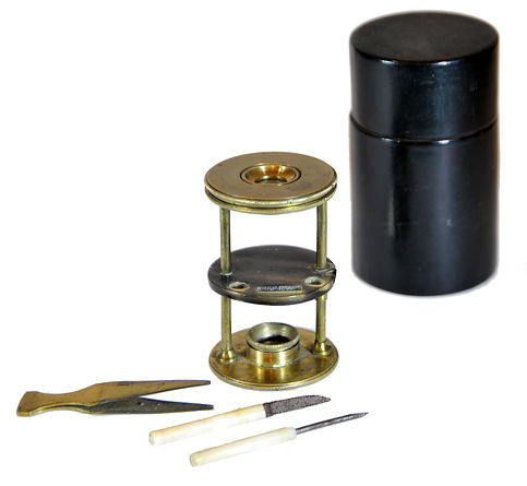

Withering-type botanical microscope, 1780
The “Withering-type Microscope” is named for its inventor, Dr. William Withering (1741-1799), an English physician and botanist who graduated with a degree in medicine 1766 in Edinburgh. Inspired by the taxonomical work and systematic classification of Carl Linnæus (1707-1778), Withering (1776) applied the Linnaean taxonomical system of classification to British plants in a seminal, two volume work, A Botanical arrangement of all the vegetables naturally growing in the British Isles. The earliest reference to a small botanical microscope of Withering’s design appeared in the first edition of this book. There, Withering indicated this microscope was developed for field dissections of flowers and other plant parts. While there is no surviving example of this exact design, close relatives of this type do exist, made either completely of brass or of ivory with brass pillars. Ivory models can be tentatively dated to 1776-1785, as by 1787 a newer model with a hollowed stage in an all-brass configuration already predominated. In turn, it was preceded by the brief appearance of a transitional brass model but with solid stage of ivory or horn (seen here). This version is extremely rare and must have been produced in very small numbers. By 1787 all these varieties were not recorded anymore in the literature.
References: SML: A242712; Goren 2014.
References: SML: A242712; Goren 2014.
Prof. Yuval Goren's Collection of the History of the Microscope

Leeuwenhoek Microscope Replica
This is a replica of the famous microscope made by Anthony Philips van Leeuwenhoek, now deposited in the Museum Boerhaave in Leiden. Of an estimated number of about 500 microscopes made by Leeuwenhoek, including 29 specimens that were sent by his daughter after his death to the Royal Society in London and later lost, only ten or eleven survived to date. One of these microscopes has an estimated magnification of 277X, but according to Brian Ford (in Single Lens, the Story of the Simple Microscope), a 500X magnification could be reached as well.
This replica was made by Christopher Allen. It magnifies ~100X.
Leeuwenhoek used such single microscopes, all made by him, for the inspection of nearly everything that he could reach, discovering on the way the blood cells, spermatozoa, bacteria, and protozoa in pond water. Together with Robert Hook, who used a compound microscope, Leeuwenhoek was the main pioneer of microscopy and one of the most significant scientists of the 17th century.
Scientific work made with this model: Leeuwenhoek used such single microscopes, all made by him, for the inspection of nearly everything that he could reach, discovering on the way the blood cells, spermatozoa, bacteria, and protozoa in pond water. Together with Robert Hook, who used a compound microscope, Leeuwenhoek was the main pioneer of microscopy and one of the most significant scientists of the 17th century.
References: Ford 1985; Boerhaave: V07017, V07018, V07019, V30337.
© Microscope History all rights reserved