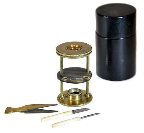

Withering-type botanical microscope, 1780
The “Withering-type Microscope” is named for its inventor, Dr. William Withering (1741-1799), an English physician and botanist who graduated with a degree in medicine 1766 in Edinburgh. Inspired by the taxonomical work and systematic classification of Carl Linnæus (1707-1778), Withering (1776) applied the Linnaean taxonomical system of classification to British plants in a seminal, two volume work, A Botanical arrangement of all the vegetables naturally growing in the British Isles. The earliest reference to a small botanical microscope of Withering’s design appeared in the first edition of this book. There, Withering indicated this microscope was developed for field dissections of flowers and other plant parts. While there is no surviving example of this exact design, close relatives of this type do exist, made either completely of brass or of ivory with brass pillars. Ivory models can be tentatively dated to 1776-1785, as by 1787 a newer model with a hollowed stage in an all-brass configuration already predominated. In turn, it was preceded by the brief appearance of a transitional brass model but with solid stage of ivory or horn (seen here). This version is extremely rare and must have been produced in very small numbers. By 1787 all these varieties were not recorded anymore in the literature.
References: SML: A242712; Goren 2014.
References: SML: A242712; Goren 2014.
Prof. Yuval Goren's Collection of the History of the Microscope


Nachet et Fils, Nachet Petite Modèle Inclinée, ca. 1875
This is the small inclining microscope by Nachet et fils, dating to circa 1875. The petite modèle was made by Nachet over a long time with several changes through its sequence of existence.
Dating the Nachet microscopes is difficult, as they bear no serial numbers. This microscope has engraved on the "Nachet et fils, 17 rue St Séverin Paris", so it was made between 1856 and 1862 when he worked at this address.
A petite modèle by Nachet was used by Louis Pasteur (1822-1895) in his studies of the principles of vaccination, microbial fermentation, and pasteurization. Pasteur used several types of microscopes throughout his career, and the microscope that he used in the early years of his research is now displayed in the Science Museum in London.
Another user of the model seen here was Santiago Ramòn y Cajal (1852-1934), the Spanish neuroscientist and pathologist, in his neuroanatomical studies of the histology of the central nervous system.
© Microscope History all rights reserved


The microscope in this collection was purchased in an antique shop in Florence, Italy. It is unprovenanced and its previous history is unknown, but by the optical components that it came with it was undoubtedly in use by a prominent scientist. The original small diameter Nachet thread for the objectives was equipped with a converter to the somewhat bigger (but not RMS standard) thread of the Hartnack optics (which were considered to be superior to those of Nachet), and the four objectives included in the set are indeed all by Hartnack. They include Hartnack's lower magnification objectives Nos. 2 and 3, but also the highly esteemed and expensive (at that time) water immersion objectives Nos. 9 and 10 with their original canisters. The higher choice than usual of oculars enable, together with the objectives, very versatile work at both lower and high magnifications with the ability to observe bacteria and other micron-scale features.
In 1859, Edmund Hartnack first exhibited his water immersion objectives (Hartley, 1993: 36, 328). He also added the correction collar to the water-immersion lens for the first time. Hartnack sold 400 of these lenses over the course of the next five years. In 1862, Hartnack displayed his immersion objectives at the London, International Exhibition. That same year Prazmowski joined Hartnack (in his Paris workshop), together they made substantial progress in the water immersion objectives, thanks to Prazmowski's combination of theory and practical skills. The result was that by the 1867 PARIS exposition, Hartnack's lenses were judged the best (Mayall Cantor Lectures: 1119). At the 1867 PARIS Exposition, Hartnack exhibited his improved water immersion objectives. The exhibit of Hartnack & Prazmowski surpassed all other entries for his new immersion lenses. That year, Hartnack produced a water-immersion objective of 1/12 inch (No.9) & 1/16 inch (No.10). These two lenses are represented here.
© Microscope History all rights reserved