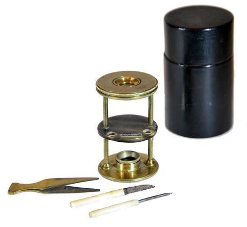

Withering-type botanical microscope, 1780
The “Withering-type Microscope” is named for its inventor, Dr. William Withering (1741-1799), an English physician and botanist who graduated with a degree in medicine 1766 in Edinburgh. Inspired by the taxonomical work and systematic classification of Carl Linnæus (1707-1778), Withering (1776) applied the Linnaean taxonomical system of classification to British plants in a seminal, two volume work, A Botanical arrangement of all the vegetables naturally growing in the British Isles. The earliest reference to a small botanical microscope of Withering’s design appeared in the first edition of this book. There, Withering indicated this microscope was developed for field dissections of flowers and other plant parts. While there is no surviving example of this exact design, close relatives of this type do exist, made either completely of brass or of ivory with brass pillars. Ivory models can be tentatively dated to 1776-1785, as by 1787 a newer model with a hollowed stage in an all-brass configuration already predominated. In turn, it was preceded by the brief appearance of a transitional brass model but with solid stage of ivory or horn (seen here). This version is extremely rare and must have been produced in very small numbers. By 1787 all these varieties were not recorded anymore in the literature.
References: SML: A242712; Goren 2014.
References: SML: A242712; Goren 2014.
Prof. Yuval Goren's Collection of the History of the Microscope

Spencer Model 60 Field Microscope, 1921
The Spencer model 60 was designed around WWI primarily for use in frontline hospitals. The weighty and thick-shelled metal carrying case is highly resistant to impact and shock. Some of these microscopes saw use in civilian laboratories, but the accessories are adequate for military service in frontline hospitals. It seems that civilian doctors were less likely to buy a 60 Model, which, in the late 1920s, had the price tag of $160 for the 60H model represented here. This was about the average monthly salary. The corresponding conventional models were by far cheaper hence more affordable.
The Spencer Model 60 is considered classic in terms of design. Therefore, it was chosen by the Microscopical Society of Southern California (MSSC) to form their emblem.


The common tendency among collectors is to include in their collections items of excellent state of preservation condition. The term "mint", which refers to an item in an as-new condition, is the aspiration of every collector. But this approach treats the object as the essence of everything and ignores its "historical patina", which constitutes the historical identity of an object, that is, its cultural and social context in time and space where it was created, and to a large extent also its provenance. I did not expect, nor do I want to get into my collection a war hero who is not scarred and bruised from the horrors of the battlefield. A veteran of the North African battles, the invasion to Normandy, the Battle of the Bulge or the Pacific Wars, will never return home unharmed and uninjured, with no bruises or changes made in the field hospital to adapt it to the harsh conditions of war. And this is also the story of the microscope before us. It carries the decoration of war on its chest, the inscription attributing it to the Medical Corps of the United States Army. But the scratches and bruises, the red re-painting of the case to highlight it and the missing items from it, are part of its historic patina and its special attraction.

