
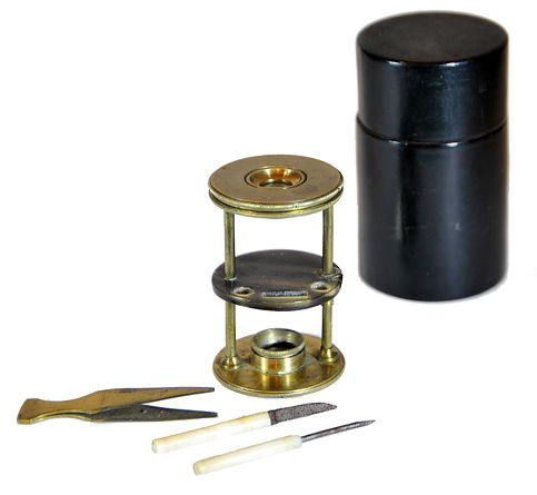
Withering-type botanical microscope, 1780
The “Withering-type Microscope” is named for its inventor, Dr. William Withering (1741-1799), an English physician and botanist who graduated with a degree in medicine 1766 in Edinburgh. Inspired by the taxonomical work and systematic classification of Carl Linnæus (1707-1778), Withering (1776) applied the Linnaean taxonomical system of classification to British plants in a seminal, two volume work, A Botanical arrangement of all the vegetables naturally growing in the British Isles. The earliest reference to a small botanical microscope of Withering’s design appeared in the first edition of this book. There, Withering indicated this microscope was developed for field dissections of flowers and other plant parts. While there is no surviving example of this exact design, close relatives of this type do exist, made either completely of brass or of ivory with brass pillars. Ivory models can be tentatively dated to 1776-1785, as by 1787 a newer model with a hollowed stage in an all-brass configuration already predominated. In turn, it was preceded by the brief appearance of a transitional brass model but with solid stage of ivory or horn (seen here). This version is extremely rare and must have been produced in very small numbers. By 1787 all these varieties were not recorded anymore in the literature.
References: SML: A242712; Goren 2014.
References: SML: A242712; Goren 2014.
Prof. Yuval Goren's Collection of the History of the Microscope
Chapter 44: The Microscope in Use by the Experts
Carl Zeiss, Jena, Stativ Va, 1882, used by Dr. Henri van Heurck
This is an early Zeiss compound microscope with the serial number 5340, dating back to 1882. The microscope, known as Zeiss Stativ Va, was originally sold to Henri van Heurck on April 24, 1882, along with four Zeiss eyepieces numbered 1, 2, 3, and 5, but without an objective. Henri-Ferdinand van Heurck (1839 - 1909), the renowned Belgian botanist and microscopist, was known for his work on diatoms and was in contact with major instrument makers in Germany, France, and the UK.


© Microscope History all rights reserved


The instrument shown here, serial number 5340, was originally sold to Dr. Henri van Heurck of Antwerp on April 24, 1882, along with four Zeiss eyepieces numbered 1, 2, 3, and 5, but without any objective. Instead, van Heurck added four objectives from the Parisian Nachet company, marked as 3, 5, 6, and 10 oil immersion, along with a magnification table on which he added his signature. All these lenses are preserved in the case. The microscope has, therefore, been preserved in its original configuration personally selected by the original owner according to the original sales records of the Carl Zeiss Archives.
The Zeiss microscope tripod Va features a fixed tube length made of lacquered and blackened or black lacquered brass, as well as blued steel. It is similar to the “Continental microscope” from the second half of the 19th century. Coarse focusing can be adjusted by sliding the optical tube through its sleeve, and fine adjustment is made using a knurled wheel on the column. It originally included a complete Abbe illumination apparatus with plane and concave mirrors, five circular aperture diaphragms, and a star diaphragm for darkfield illumination. As an alternative to Abbe's illumination apparatus, a simple apparatus was included, which consists of a five-bearing mirror for movement outside the optical axis and the appropriate cylinder diaphragm on slides.

In Zeiss catalogue No. 25 (1881): Illustrated catalogue of microscopes and auxiliary equipment from the optical workshop of Carl Zeiss in Jena, this model appears together with the optional “No. 38 revolver for four objectives, with the narrow thread of Zeiss objectives…" and "No. 66 Illumination apparatus according to Abbe condenser with a large aperture, with diaphragm apparatus and double mirror; for all… only suitable for the larger stands from I to Va”. Undoubtedly van Heurck gave up this apparatus, as well as the Abbe condenser (fit for the Zeiss optics) because he preferred the objectives made by the Nachet company that these accessories wouldn’t fit.


All this testifies to Van Heurck as a professional microscopist who customized his microscope to fit his specific needs by combining products from different top European manufacturers. His expertise is evident in his famous book The Microscope and in the later development of the high-quality microscope model that was named after him by the British Watson Company, produced from the late 19th century through the first decades of the 20th century.
Van Heurk and the diatoms
A. van Leeuwenhoek observed diatoms as early as 1702. The first taxonomy of diatoms was created in the Netherlands by R. B. van den Bosch in 1846 and in Flanders by J.-J. Kickx in 1867. By the second half of the nineteenth century, diatoms were already being used in geological research. H. van Heurck's "Synopsis," published between 1880 and 1885, enabled many twentieth-century researchers to conduct applied studies for geological and ecological purposes. Henri Van Heurck's book, first published in 1896, begins with an introduction to the structure, life history, and classification of diatoms. It includes detailed instructions on how to collect, cultivate, and prepare diatoms for study. The author also provides a comprehensive overview of various methods and tools used in diatom analysis, including microscopy, staining, and chemical treatments. The book is richly illustrated with detailed drawings and photographs of diatoms, along with examples of their habitats and environments. This seminal work remains an important reference for anyone interested in studying diatoms, their ecology, and their role in the natural world.
© Microscope History all rights reserved
Carl Zeiss, Stativ Ic, 1903

This microscope was manufactured in 1903. It is based on a new design by Max Berger of Zeiss, first introduced in 1898 (Zeitschrift fur Instrumentenkunde, vol. XVIII, 1898, pp. 129-133). This microscope bears the serial number 37910, dating it to 1903. The major innovations of this design were a new type of fine adjustment and a limb having an integral handle (now, often referred to as a "Jug-Handle" microscope). While this microscope is perfectly suited for conventional work, this particular model was made for the purposes of photomicrography and projection. Accordingly, the main tube and the knobs are made of aluminum, presumably to reduce the weight on the fine adjustment mechanism, thus allowing it to be more sensitive and responsive when the microscope is inclined in the horizontal position (later versions of this model dispensed with the use of aluminum). (Article from the Quekett Microscopical Club).
The plan and concave mirrors are here still connected via a dovetail with the carriage of the lighting apparatus. Later tripods of this type are characterized by an inserted into the end of the rack mirror fork. A large cross table takes up the rehearsal. This microscope stand has been offered since 1898 as the largest and for all microscopic work suitable tripod. Due to the large tube diameter, the microscope is very well suited for micro-projection and photomicrography. Funnels added to the tripod allow easy mounting of a camera.
Carl Zeiss Photo-Micrographic Microscope - Carl Zeiss Jena produced a range of photo-micrographic microscopes designed for use in photography and microscopy, with the ability to capture high-quality images of microscopic specimens. One notable example is the Carl Zeiss Jena Photo-Micrographic Microscope or Ph Stand, which was a popular model in the early to mid-20th century. This microscope was designed for use in photomicrography, which involves taking photographs of microscopic specimens using a microscope and a camera. It was known for its high-quality optics, which allowed for clear and precise imaging of specimens. It also featured a range of adjustable controls and accessories to customize the microscope for different types of specimens and imaging needs.
References: Mappes; German Museum Munich, Inv.-No. 3453; Collection of the Royal Microscopic Society, Inventory no. 272: 310 and 319; Billings Collection Washington, AFIP 17776 - 60-4713-107, p. 120, Fig. 226, and AFIP 39-60-4713-385, p. 123, Fig. 234; The Microscope Collection at the Science Museum London, Inventory No. 1989-151, 1986-528 and 1992-1094; Optical Museum of the Ernst-Abbe-Foundation Jena; Optical Museum Oberkochen; Pathological-anatomical Federal Museum Vienna, Museum Nos. 25.346, 29,093 and 26,568; Microscope Collection of the Moscow Polytechnic Museum, inventory number PM 007897 (MIM 430), Oxford Museum of History of History, Inventory Nos. 20525 and 68970; Boerhaave Museum Leiden, NL, inventory # V03062; Historic Microscopes at the Laupus Health Sciences Library, East Carolina University, Inventory no. G4

Carl Zeiss, "Bierseidel" Stativ IIIE, 1909
While this collection is focusing on pre-20th century microscopes, it was decided to include in it this very fine example of a transitional 19th-20th century microscope by the then (and now) world-leading optical company of Carl Zeiss, Germany.
The profile of this instrument features an integrated “jug handle” (German: "Bierseidel"), cut into a squared off limb, a design that became popular among some producers between ca. 1900 and 1920. The serial number, 43430, indicates the production year 1909. Typical of microscopes of the turn of the 19th century to about 1930 is the combination of original lacquered brass tube, a remnant of the 19th century, with the black-painted ("japanned") limb and horseshoe base, to be replaced by the all-black body during the later decades of the 20th century.
Having already all the optical advances of the second half of the 19th century, scientifically set by Ernst Abbe at the initiative of Carl Zeiss, together with the advanced form which developed from the "Continental stand" of the earlier decades, this is a fully capable research instrument. Even today, over 110 years later, this instrument can still produce crisp and perfect views at any magnification between 50x and 1000x.
